Label-free Multiphoton Imaging
Two-Photon Morphometry in Gut and Bowel Disease
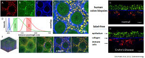
In collaboration with the Erlangen Medical Clinics I (Gastroenterology), we apply our two-photon imaging and morphometry approach to human bowel biopsy samples obtained during colonoscopy to study structural tissue architecture remodeling in inflammatory bowel disease (IBD). Applying spectral separation of optical two-photon-excited autofluorescene (AF) and SHG signals, we can structurally visualize molecular non-linear optical fingerprints of collagen-I (extracellular matrix), NADH (enriched in epithelial cells) and FAD (enriched in immune cells), see figure below (A-F). The figure shows spectrally separated images from one fresh human colon biopsy in depth through the various layers of epithelium, lamina propria and submucosa containing immune cells (F). G, and H, show side views obtained from XYZ image stacks from a fresh human colon biopsy sample from a healthy person and a patient with Crohn’s disease, respectively. Within the inflamed submucosa, a marked immune cell infiltration can be observed. We are currently developing automated image processing pattern recognition algorithms to facilitate IBD diagnsotics including multiphoton imaging modalities.
 Morphological representation of colon mucosa architecture utilizing two-photon induced autofluorescence (NADH, FAD) and SHG signals that can be excited with one wavelength and emission spectrally separated with optical filters. A, FAD channel representing immune cells, B, NADH channel representing epithelial cells and C, SHG channel representing extracellular matrix alongside with overlay and spectral bins (D). F, sections through an XYZ stacks through the biopsy sample. G, side view reconstructed from XYZ stack of a healthy sample and a human patient with Crohn’s disease (H) showing marked immune cell infiltration (Schürmann et al. 2013).
References:
- Schürmann S, Foersch S, Atreya R, Neumann H, et al. (2013) Label-free imaging of inflammatory bowel disease using multiphoton microscopy. Gastroenterology. 45(3):514-6. doi: 10.1053/j.gastro.2013.06.054.
